

Highlights
-
•
“Total situs inversus” is an infrequent congenital condition.
-
•
The robot has been already proved as a safe and attractive approach for living donor neprectomies.
-
•
“Total situs inversus” presents the surgeon with a challenging situation.
-
•
In these cases, preoperative planning is essential.
-
•
This is the first robotic right donor nephrectomy in a patient with total sinus inversus.
Keywords: Living donor, Transplant, Situs inversus, Robotic nephrectomy, Hand-assisted, Kidney
Abstract
Total situs inversus” is an infrequent congenital condition. The robot has been already proved as a safe and attractive approach for living donor neprectomies. We report here the first right donor nephrectomy in a patient with total sinus inversus that is performed using the Da Vinci platform.
1. Introduction
“Total situs inversus” is an infrequent congenital condition with a prevalence of 0.01% of the population that involves transposition through the saggital plane of all internal organs [1]. It has been considered an absolute contraindication for liver and heart transplantation and it poses the surgeon to a challenge situation. However there have been reported TSI in donor nephrectomies. The robotic platform has been already proved as a safe and attractive approach for the donor nephectomies with a decreased in the length of stay [2]. This is the first report of a right donor nephrectomy in a patient with total sinus inversus that is performed using the Da Vinci platform.
2. Patient and methods
Patient is a 34 year-old African American female with BMI of 29.2 kg/m2 no past medical or surgical history, presenting as a leaving donor for her relative for kidney transplantation. The preoperative imaging computed tomography revealed a “total situs inversus” with the right kidney with a single long right renal vein draining into a left-sided vena cava and a single short right renal artery behind the renal vein (Fig. 1). Decision was made to perform a right nephrectomy to take advantage of the longer renal vein on the right side in this particular anatomic situation (Fig. 2).
Fig. 1.
CT Axial view: “Total Situs Inversus” demonstrating the right renal vein draining into the left-sided Vena Cava.
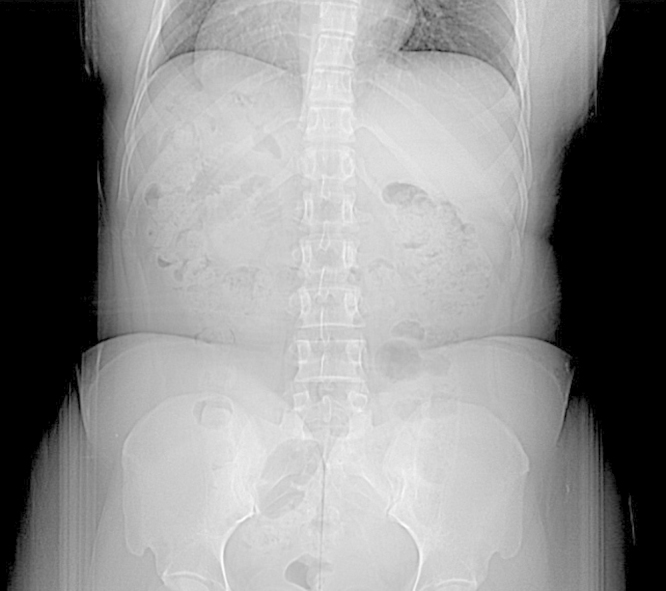
Fig. 2.

CT Coronal view: “Total Situs Inversus” with the apex of the heart and stomach on the right, and liver on the left.
3. Surgical technique
The patient was positioned left lateral decubitus exposing the right flank.
A short 8 cm midline infraumbilical incision was made, the lap-disc device was then inserted into the abdominal cavity and secured. A supraumbilical camera port is introduced in the abdomen, with the assistant’s hand placed into the abdominal cavity protecting the underlying viscera. An additional 8 mm robotic trocar was placed in the mid clavicular line superior to the camera port, another 8 mm trocar was placed at the left of the camera trocar, and a 12 mm trocar in the lower right quadrant, and finally the arms of the robot were attached to the trocars.
The assistant’s right hand was introduced into the abdominal cavity via the hand assisted window and used to expose the left colic gutter by providing counter tension on the left descending colon located on the patient’s right side. The kidney in its retroperitoneal position was readily exposed after medial rotation of the colon. The right ureter was readily identified at the pelvic brim and circumferentially dissected along with the gonadal vein using a combination of sharp instrument and then finger dissection. Care was taken to not disturb the vascular supply to the ureter or the fat surrounding it. Once isolated, a ½ inch penrose drain is looped around the ureter and gonadal facilitating atraumatic retraction of the ureter lateral and anteriorly. In following the ureter/gonadal complex superiorly, we were able to readily identify the right renal vein.
Gerota’s fascia was incised superiorly and the kidney surface identified, developing a plane between the superior portion of the kidney and the adrenal gland. The renal vein was circumferentially dissected using the hook cautery. The gonadal and adrenal veins are dissected and divided. The kidney was then mobilized posteriorly by dividing its loose areolar attachments exposing the back to the hilum of the kidney. Two posterior lumbar veins were dissected out and then doubly clipped in continuity and divided. The posterior aspect of the renal vein was then extensively freed up by dividing small lymphatics surrounding it until there was sufficient length. Next, the renal artery was circumferentially isolated by dividing overlying nerves and lymphatics using the hook cautery. The artery was dissected all the way back to its take off from the aorta.
Once the vascular pedicle is prepared, the ureter was divided using clips. Systemic heparinization is administered and a vascular TA stapler was used to divide the renal artery, secured with a hemolock and transected with robotic scissors. The kidney in rotated to its anatomical position and the renal vein is divided with an endoGIA with vascular load. The kidney was immediately removed by the assistant and carried to the back Table where it was reperfused with cold Wisconsin’s solution. The total warm ischemia time was less than 90 s. Hemostasia is obtained and closure by layers is completed. The operative time was 170 min including docking time. Blood loss was minimal. There were no perioperative or post-operative complications. The post-operative evolution was uneventful and patient was discharged home on the third post-operative day.
4. Discussion
“Total situs inversus” is an rare congenital condition [1]. Orientation is more complicated and surgery wise it is a challenge for the surgeon, however it has not been cited as exclusion criteria. Multiples procedures has been reported in STI, but not many in living donor nephrectomy. It is a challenge situation for the surgeon that has to adjust the trocars placement to the anatomy target location in a minimal invasive approach. In these cases preoperative studies are essential in order to devise surgical approach and minimize risks. In a meta-analysis published recently by Wang K, paper showed similar effect on the surgery and postoperative graft function in right and left laparoscopic living-donor nephrectomy, however the longer renal vein of the left kidney could decrease operative difficulty and it was recommended [3]. In this case, decision was made to use the right kidney for the longer renal vein due to the atypical anatomical conformation. The procedure is performed following similar steps to patients with normal anatomy, there was no particular anatomical differences to the commonly performed left nephrectomy but mirror image. There is scarce literature regarding this topic. A paper by Berber and colleagues reported a case where the donor nephrectomy in a situs inversus was performed using a total laparoscopic technique [4]. To our knowledge, this is the first donor nephrectomy performed in a patient with situs inversus using the robotic platform. The endowrist technology and the HD, 3D camera of the Da Vinci system, provide the surgeon an improved vision and precise dissection. The robotic technology is safe and facilitates the procedure in presence of the most common anatomical variations, those are multiple arteries and less frecuently a retroaortic left renal vein [5]. This particular anatomical variation should not be considered a contraindication for organ donation.
5. Disclosure
The authors of this manuscript have no conflicts of interest to disclose.
Conflict of interest
Authors have nothing to disclose.
Funding
No funding.
Consent
Patients consented for research.
Author contribution
Raquel Gonzalez-Heredia, MD/PhD: writing and editing the manuscript Raquel Garcia-Roca, MD: writing and editing the manuscript Enrico Benedetti, MD, Head of Department of Surgery: final edition of the manuscript.
Guarantor
Enrico Benedetti, MD, Head of Department of Surgery.
Contributor Information
Raquel Gonzalez-Heredia, Email: rgheredi@uic.edu.
Raquel Garcia-Roca, Email: raqgar@hotmail.com.
Enrico Benedetti, Email: enrico@uic.edu.
References
- 1.Nursal T.Z. Laparoscopic cholecystectomy in a patient with situs inversus totalis. J. Laparoendosc. Adv. Surg. Tech. A. 2001;11(4):239–241. doi: 10.1089/109264201750539772. [DOI] [PubMed] [Google Scholar]
- 2.Cohen A.J. Robotic-assisted laparoscopic donor nephrectomy: decreasing length of stay. Ochsner J. 2015;15(1):19–24. [PMC free article] [PubMed] [Google Scholar]
- 3.Wang K. Right versus left laparoscopic living-donor nephrectomy: a meta-analysis. Exp. Clin. Transplant. 2015;13(3):214–226. [PubMed] [Google Scholar]
- 4.Berber I. Total laparoscopic donor nephrectomy in situs inversus totalis: a case report. Exp. Clin. Transplant. 2013;11(2):195–198. doi: 10.6002/ect.2012.0098. [DOI] [PubMed] [Google Scholar]
- 5.Gorodner V. Routine left robotic-assisted laparoscopic donor nephrectomy is safe and effective regardless of the presence of vascular anomalies. Transpl. Int. 2006;19(August (8)):636–640. doi: 10.1111/j.1432-2277.2006.00315.x. [DOI] [PubMed] [Google Scholar]