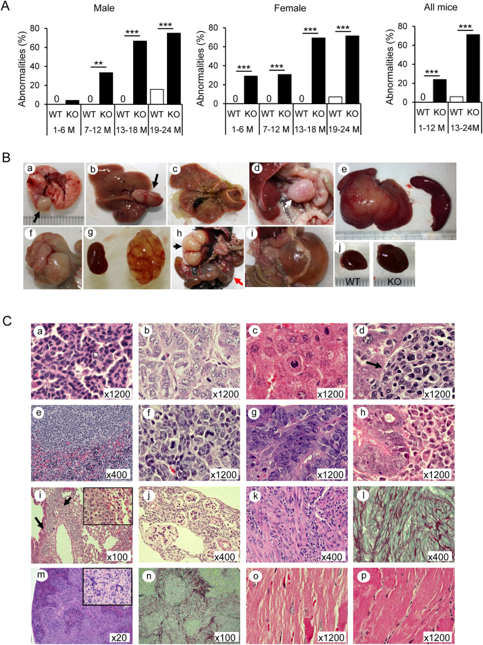
Figure 1. Multiple organ abnormalities in Cygb−/− mice.
(A) Percentage of abnormalities found in male (left panel), female (middle panel), and all (right panel) wild-type (WT) and Cygb−/− (KO, knockout) mice in four age groups: 1–6 months of age (1–6 M), 7–12 months (7–12 M), 13–18 months (13–18 M), and 19–24 months (19–24 M). Open bar, WT; closed bar, KO. Data represent the mean ± SD; n = 15–56 per group; **p < 0.01; ***p < 0.001. (B) Macroscopic findings in Cygb−/− mice: tumour nodule (black arrow) of the lung at 18 M (a); liver tumour, 17 M (b); liver cholestasis, 5 M (c); swelling of mesenteric lymph node, 23 M (d, white arrow); hepatosplenomegaly, 11 M (e); intestinal tumour, 24 M (f); kidney cyst, 4 M (g); kidney deformity (black arrow) and cyst of uterus (red arrow), 6 M; mesenteric cyst, 10 M (i); heart hypertrophy, 22 M (j). (C) Representative haematoxylin and eosin (H&E)-stained sections of Cygb−/− mice with adenoma (a) and adenocarcinoma (b) of the lung; hepatocellular carcinoma (HCC) (c); lymphoma in the liver (d, arrow), spleen (e), and mesenteric lymph node (f); intestinal adenoma (g); intestine lymphoma (h), which was metastatic to the lung (i, arrows; right inset, ×1200); kidney cysts (j); H&E and Sirius Red and Fast Green (SiR-FG) staining of a renal myomatous lesion (k, l) and spleen fibrosis (m, n; right inset, ×800); hypertrophy of cardiomyocytes in KO mice compared with WT mice (o, WT; p, KO).

