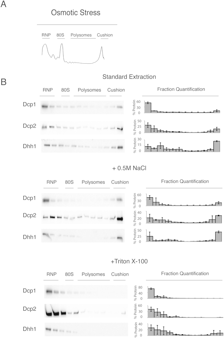
Figure 4. During osmotic stress, the decapping factors were localised to the rapidly sedimenting/dense fractions in the sucrose gradient.
(A) Typical sucrose gradient profile (at 254 nm) of extracts from mid-log phase of growth, after osmotic stress (15’ treatment with 1M KCl). The extracts were separated on a 15–50% sucrose gradient. The trace demonstrates the RNP, ribosomal peaks (40S, 60S, 80S), polysomal peaks and the peak of the sucrose cushion. (B) Western blot analysis of the fractions from the above condition. To the right of the blots is the percentage of protein found in each fraction; error = SD; n = 3. The middle and bottom panels show the tagged mRNA decapping proteins from cells after lysis with 0.5M NaCl or with buffer containing 1% Triton X-100. The localisation of the RNP, 40S, 80S, polysome and cushion regions of the gradient is noted above the western blots.

