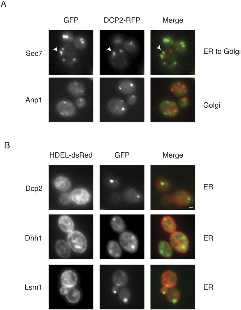
Figure 8. The decapping factors are adjacent or co-localised with the Golgi apparatus and/or ER.
(A) The exponential-phase cells that expressed chromosomal GFP-tagged proteins (indicated to the left) that expressed Dcp2-RFP on a plasmid were glucose deprived for 15 minutes. The GFP-tagged protein localisation is indicated to the right. An arrow indicates the adjacent Dcp2 and Sec7 foci; the scale bar is 1 μm. (B) Exponential-phase cells that expressed both chromosomal GFP-tagged decapping proteins (indicated to the left) and chromosomally integrated HDEL-dsRed were subjected to 15 minutes of glucose deprivation. The localisation of HDEL-dsRed is indicated to the right; the scale bar is 1 μm.

