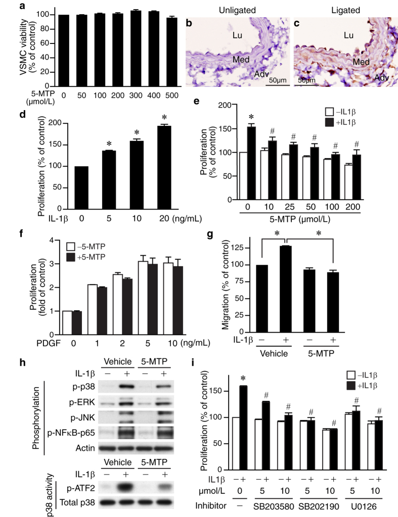
Figure 4. 5-MTP suppresses VSMC proliferation via p38 MAPK and ERK pathway.
(a) MTT assays were performed to assess the effects of 5-MTP on VSMC viability. Immunohistochemistry of (b) unligated and (c) 4 d-ligated vessel sections were performed to detect IL-1β expression (brown). (d) Serum-starved VSMCs were treated with different doses of IL-1β for 24 h, proliferation then assessed by BrdU incorporations and normalized to control without IL-1β stimulation. *P < 0.05 vs. control. (e) Serum-starved VSMCs (in the absence or presence of different doses of 5-MTP) were treated with IL-1β for 24 h and proliferation assessed and normalized to control without IL-1β stimulation. *P < 0.05 vs. control; #P < 0.05 vs. IL-1β-treated but without 5-MTP. (f) Serum-starved VSMCs were stimulated with different concentrations of PDGF-BB in the presence or absence of 5-MTP (100 μmol/L) for 24 h and proliferation assessed. (g) Serum-starved VSMCs (in the absence or presence of 5-MTP) were treated with IL-1β for 24 h, and migration assays performed. VSMCs migrating through the filters were quantified after 4 h. *P < 0.05. (h) Upper panel, VSMCs were serum starved in the absence or presence of 5-MTP, stimulated with or without IL-1β for 15 min, and total proteins isolated for Western blotting to detect phosphorylations of p38 MAPK, ERK1/2, JNK, and NFκB-p65 (Ser536). To verify equal loading, the blots were probed with a pan-actin antibody. A representative of 3 independent experiments is shown. Lower panel, p38 MAPK activity of VSMCs was measured using a p38 MAP kinase assay kit. Phosphorylation of ATF2 indicates p38 MAPK activity. Aliquots of cell lysates were subjected to Western blotting with p38 antibody to verify equivalent amount of protein for activity assessment. A representative of 3 independent experiments is shown. (i) VSMCs were pretreated with p38 MAPK inhibitor SB203580 or SB202190, or MEK inhibitor U0126 30 min prior to IL-1β stimulation. Proliferation was then assessed 24 h later. *P < 0.05 vs. control without IL-1β; #P < 0.05 vs. IL-1β-treated but without inhibitor. Values are mean ± SE of at least three experiments.

