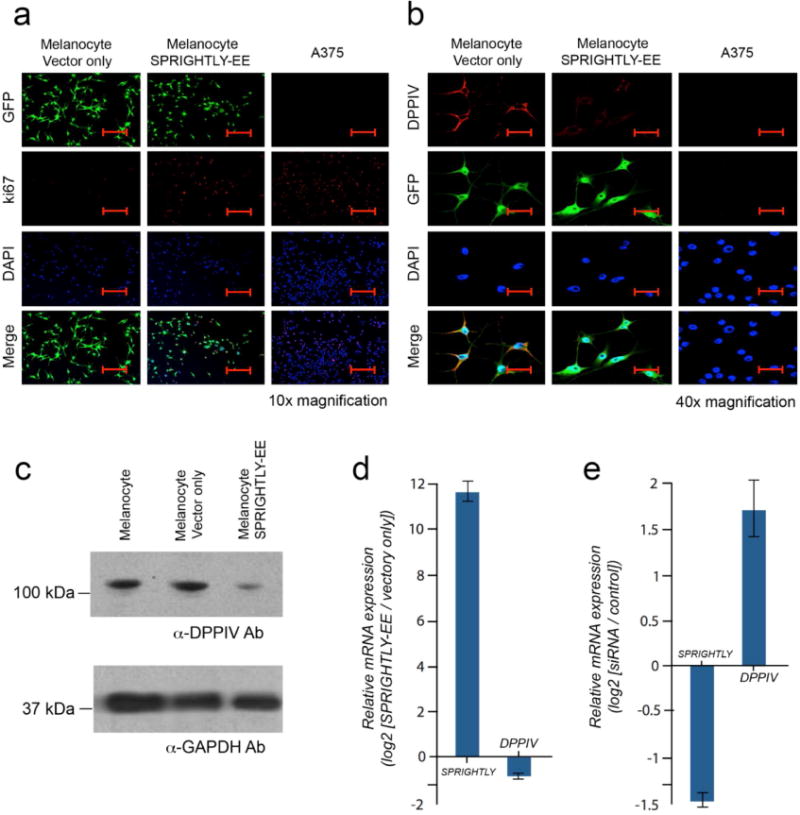
Figure 5. Ki-67 and DPPIV expression in melanocytes ectopically expressing SPRIGHTLY.

(a) Immunofluorescence staining of Ki-67 in SPRIGHTLY-EE, Vector-only melanocytes, and A375 cells. Ki-67 nuclear staining is highest in A375 cells. Ki-67 staining is elevated in SPRIGHTLY-EE cells compared to Vector-only controls. Scale bar = 80 um. (b) Immunofluorescence staining of DPPIV in SPRIGHTLY-EE cells, Vector-only melanocytes, and A375 cells. Control melanocytes display abundant cell-surface DPPIV, A375 cells express little or no DPPIV, and SPRIGHTLY-EE cells show intermediate expression levels, i.e., an inverse pattern to Ki-67. Scale bar = 20 um. (c) DPPIV protein levels (western blot) in SPRIGHTLY-EE cells relative to melanocytes alone and melanocytes transfected with empty vector. (d) Ectopic expression of SPRIGHTLY downregulates DPPIV mRNA levels in melanocytes. (e) siRNA-mediated knockdown of SPRIGHTLY upregulates DPPIV mRNA levels in A375 melanoma cells.
