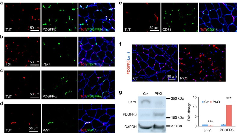
Figure 2. Specificity of Pdgfrβ-driven Cre.
(a–e) TdT (red) expression co-localized with PDGFRβ (a, green), PW1 (d, green), but not Pax7 (b, green), PDGFRα (c, green) or CD31 (e, green) in Ai14:Pdgfrβ-Cre+ reporter mice. TdT, tdTomato. (f) Laminin γ1 (blue) and PDGFRβ (red) expression in tibialis anterior muscles. (g) Western blot analysis of laminin γ1 and PDGFRβ expression in skeletal muscles. GAPDH was used as a loading control; n=4. Scale bars, 50 μm. ***P<0.001 (Student's t-test). The results are shown as mean±s.d.

