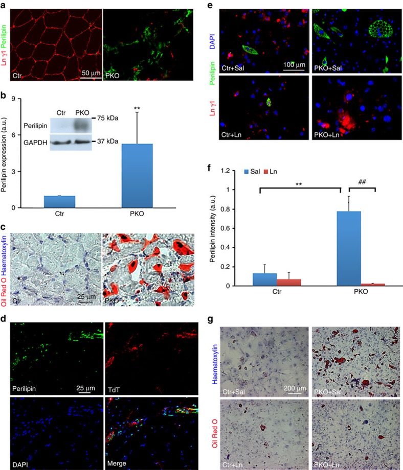
Figure 6. Laminin inhibits the adipogenesis of PDGFRβ+ cells.
(a) Perilipin (green) and laminin γ1 (red) expression in hindlimb muscles from 2-month-old mice. (b) Western blot analysis of perilipin expression in skeletal muscles. GAPDH was used as a loading control; n=6. (c) Oil Red O staining of hindlimb muscles from 2-month-old mice. (d) Perilipin (green) expression co-localized with TdT (red) in the muscles of laminin-deficient reporter (F/F:Ai14:Pdgfrβ-Cre+) mice. (e) Perilipin (green) and laminin γ1 (red) expression in control and PKO PDGFRβ+ cells after 20 days in adipogenic differentiation in the presence of saline or exogenous laminin. (f) Quantification of perilipin expression in e; n=3. (g) Oil Red O and haematoxylin staining of primary PDGFRβ+ cells after adipogenic differentiation. Scale bars, 50 μm in a, 25 μm in c and d, 100 μm in e and 200 μm in g. **P<0.01; ##P<0.01 (Student's t-test). The results are shown as mean±s.d.

