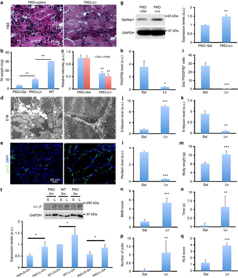
Figure 8. Exogenous laminin partially rescues MD phenotype in PKO mice.
(a) H&E staining of hindlimb muscles from PKO mice after saline or laminin injection for 2 months. (b) Quantification of tibialis anterior muscle weight; n=4. (c) Quantification of CD3 and F4/80 expression in PKO muscles after saline (Sal) or laminin-111 (Ln) treatment for 2 months; n=3. (d) Representative ultrastructural images of PKO muscles after saline and laminin injection. BM, basement membrane; #, gap. (e) Laminin γ1 (green) expression in PKO muscles after saline or laminin injection for 2 months. (f) Western blot analysis of laminin γ1 expression in wild-type (WT) and PKO muscles after saline (S) or laminin-111 (L) treatment for 2 weeks (2w) or 2 months (2m); n=3. (g) Western blot analysis of gpihbp1 expression in PDGFRβ+ cells isolated from saline- or laminin-treated PKO mice; n=3. (h,i) Quantification of PDGFRβ expression (h) and PDGFRβ+Edu+ cells (i) in PKO mice after treatment; n=3. (j,k) Quantification of S-Myosin (j) and E-Myosin (k) expression in PKO mice after treatment; n=3. (l) Quantification of perilipin expression in PKO mice after treatment; n=3. (m–q) Quantification of body weight (m), BMS score (n), suspension time (o), number of pulls (p) and HLS score (q) in PKO mice treated bilaterally with saline or laminin for 2 months; n=8. Scale bars, 25 μm in a, 2 μm in d and 50 μm in e. *P<0.05; **P<0.01; ***P<0.001 (Student's t-test). The results are shown as mean±s.d.

