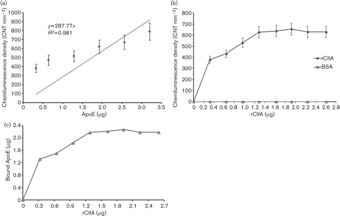
Fig. 4.
(a) Calibration curve of human ApoE (0.32–3.2 µg) dot blotted on PVDF membrane and probed for ApoE. (b) Serum ApoE binding to dot blotted rClfA. Density of the chemiluminescence signals of ApoE in diluted human serum (20 %, v/v, human serum in TBS–BSA) that bound to escalating concentrations of rClfA (0.32–2.6 µg) on PVDF membrane (BSA was used as a control). (c) Calculated ApoE (µg) in diluted human serum (20 %, v/v, human serum in TBS–BSA) that bound to escalating concentrations of rClfA (0.32–2.6 µg) on PVDF membrane using immunodot-blot assay.

