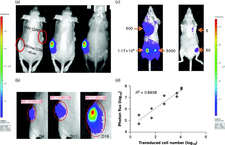
Fig. 3.
In vivo imaging of SIVmac239-eLuc-infected cells based on eLuc. (a) BLI was performed on day 11 post-transplantation and overlaid onto a photographic or X-ray image to visualize the transplanted HCT116 cells transduced with SIVmac239-eLuc. (b) The progression of tumours in the same mice was monitored in real-time by BLI. (c) In total, 5 single cells to 1.1 × 10⁴ cells infected with SIVmac239-eLuc were transplanted subcutaneously in mice and imaged by BLI 36 h post-transplantation. (d) The number of SIVmac239-eLuc-transduced cells was plotted against total photon flux emission.

