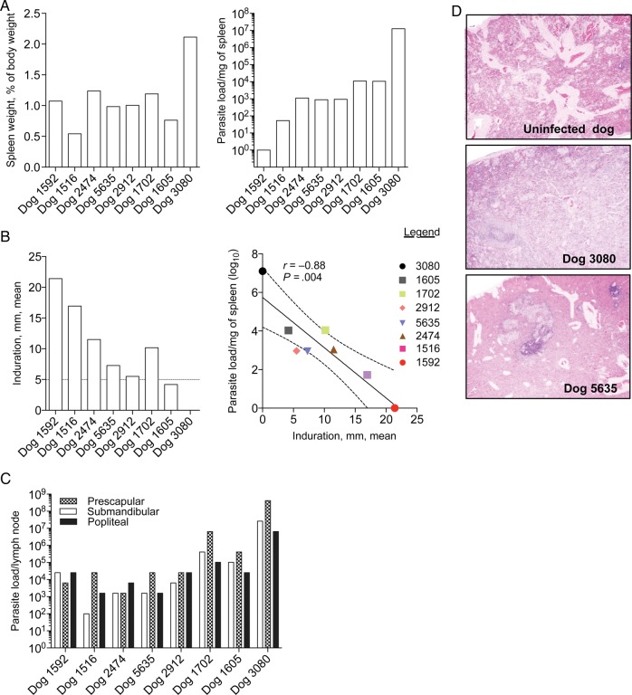
Figure 2.
Pathology and leishmanin test reactions 22 months after experimental vector transmission to beagles. A, Spleen weight as a percentage of body weight (left) and parasite load in homogenized spleen tissue (right). B, The left panel shows leishmanin test indurations measured 48 hours after intradermal injection of soluble Leishmania antigen prior to necropsy. The mean of 2 perpendicular measurements delimiting the induration area is given. A mean induration of ≥5 mm (dotted line) was considered positive. The right panel shows a negative correlation between splenic parasite load and leishmanin test positivity. The solid line represents the mean. C, Parasite loads per lymph node for submandibular, prescapular, and popliteal lymph nodes. Parasite loads were determined by a limiting dilution assay. D, Hematoxylin and eosin–stained sections of the most affected part of the spleen in representative dogs 3080 and 5635. A section from the spleen of an uninfected dog was used as control. Images were taken at the magnification ×40.

