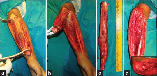
Figure 3.

Intra-operative picturing showing (a and b) mobilization of the distal ulna and oblique olecranon osteotomy at the base of the coronoid process. (c) Excised specimen. (d) Fixation of the radius to the remaining olecrenon with compression screw and tension band wiring
