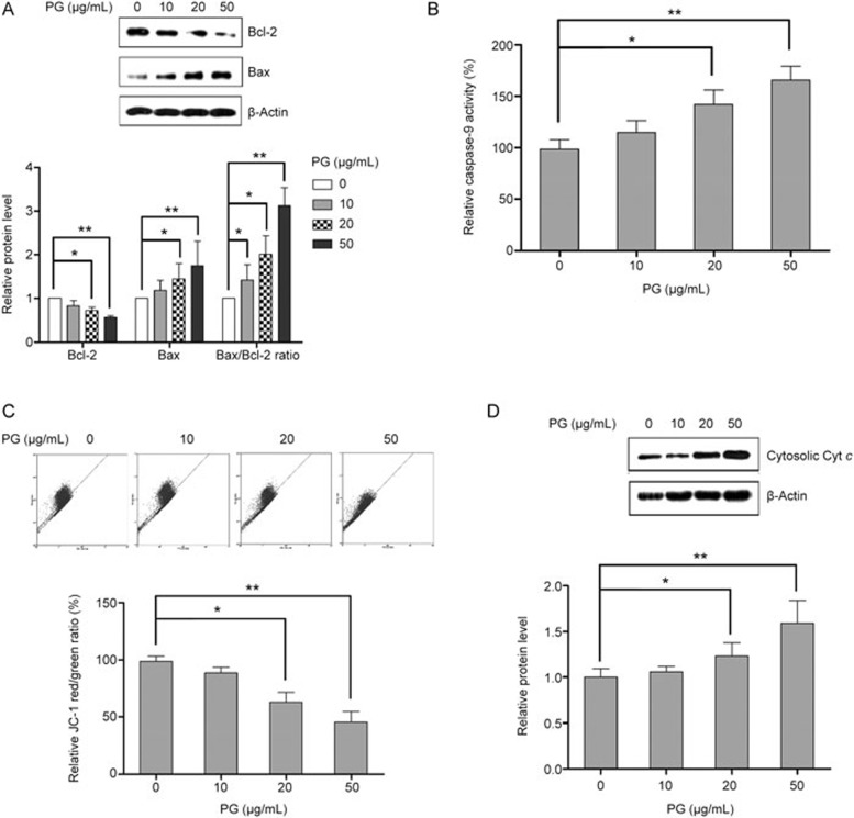
Figure 2.
PG induces apoptosis in KB cells via the mitochondrial pathway. KB cells were challenged with PG at different doses for 48 h. (A)PG increases the Bax/Bcl-2 ratio in a concentration-dependent manner. (B) PG activates caspase-9 in a concentration-dependent manner. (C) PG causes the loss of mitochondrial membrane potential. (D) PG treatment results in an increase in cytosolic cyt c. Mean±SD. n=3. *P<0.05, **P<0.01.

