Figure 6.
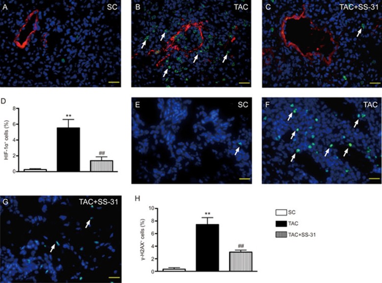
Immunofluorescent (IF) staining for identifying the expression levels of HIF-1α+ and γ-H2AX+ cells in lung parenchyma at day 60 after the TAC procedure. (A–C) IF microscopic findings for the identification of positively stained hypoxic inducible factor (HIF)-1α cells (white arrows) in lung parenchyma (×400). (D) Analytic results of the number of HIF-1α+ cells. (E–G) IF microscopic findings for the identification of positively stained γ-H2AX cells (white arrows) in lung parenchyma (×400). (H) Analytic results of the number of γ-H2AX+ cells. The scale bars in the right lower corner represent 20 μm. All statistical analyses were performed by one-way ANOVA followed by Bonferroni multiple comparison post hoc test. SC = sham control. TAC = transverse aortic constriction. Mean±SEM. n=8 for each group. **P<0.01 vs SC group. ##P<0.01 vs TAC group.

