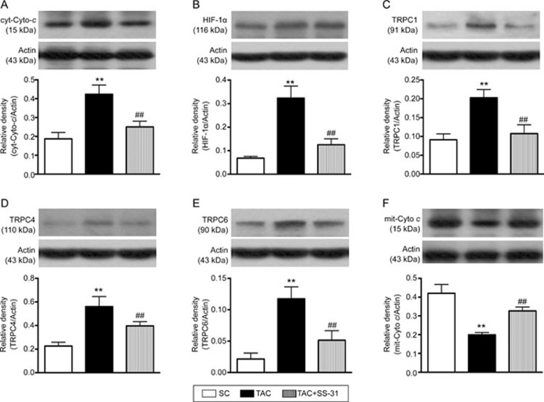
Figure 8.
Protein expression levels of mitochondrial damaged and hypoxia-induced smooth muscle proliferation markers in lung parenchyma at day 60 after the TAC procedure. (A) Protein expression of cytosolic cytochrome c(cyt-Cyto c). (B) Protein expression of hypoxic inducible factor (HIF)-1α. (C) Protein expression of transient receptor potential cation channel 1 (TRPC1). (D) Protein expression of TRPC4. (E) Protein expression of TRPC6. (F) Protein expression of mitochondrial cytochrome c (mit-Cyto c). All statistical analyses were performed by one-way ANOVA followed by Bonferroni multiple comparison post hoc test. SC=sham control. TAC=transverse aortic constriction. Mean±SEM. n=8 for each group. **P<0.01 vs SC group. ##P<0.01 vs TAC group.

