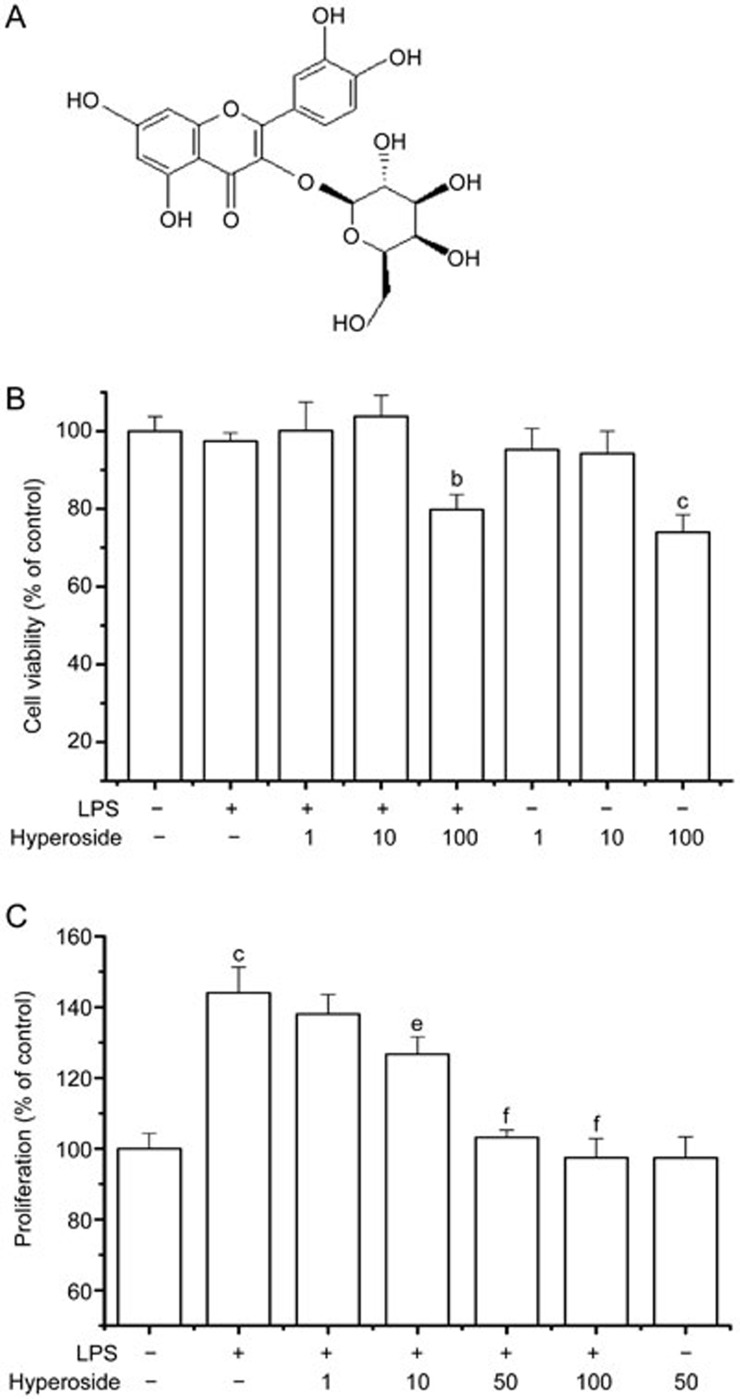
Figure 1.
Effects of hyperoside on LPS-induced RA FLSs proliferation. (A) Chemical structure of hyperoside. (B) FLSs were incubated with the indicated concentrations of hyperoside in a serum-free medium for 48 h, and cell viability was measured by the MTT assay. (C) RA FLSs were pretreated with hyperoside (1, 10, 50, and 100 μmol/L) for 1 h and exposed to LPS (1 μg/mL) for 48 h. Cellular proliferation was measured using the BrdU incorporation assay. Each value represents the mean±SEM of the three independent experiments. *P<0.05, **P<0.01 compared with the control group. #P<0.05, ##P<0.01 compared with the LPS-treated group.

