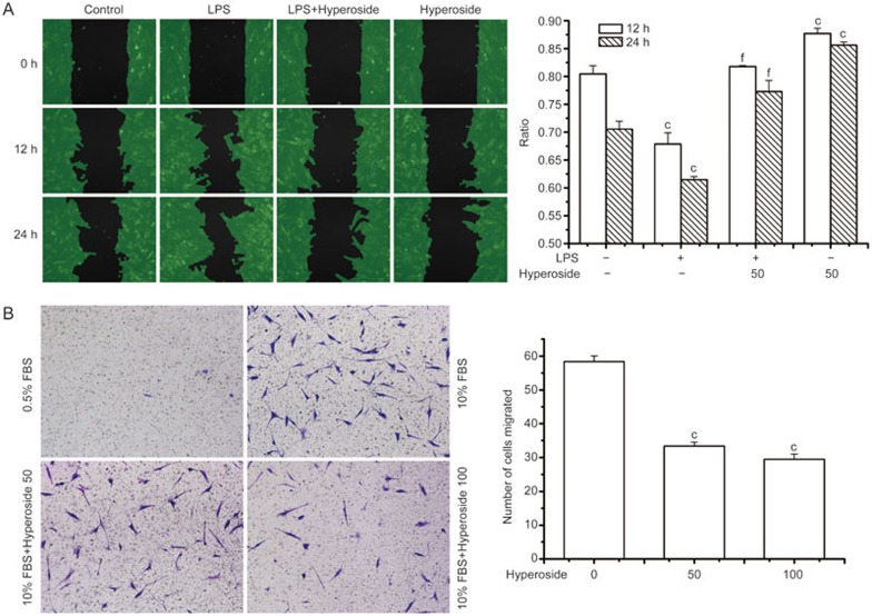
Figure 2.
Effect of hyperoside on RA FLSs migration. (A) Migration analysis using wound assay chambers. RA FLSs were seeded into wound assay culture inserts (ibidi) and incubated for 24 h. Removal of the insert yielded a uniform “wound” to the monolayer. After preincubation with hyperoside (50 μmol/L) for 1 h, cells were stimulated with LPS (1 μg/mL) for 24 h. The cell-free areas were measured using Wimasis Image Analysis (ibidi) and the ratio was calculated (cell-free area in 12 h or 24 h to cell-free area in 0 h). (B) Migration was performed in a Transwell chamber and chemotaxis was quantified by counting the migrated cells. The RA FLSs were seeded in a Transwell chamber and allowed to migrate for a further 24 h. FBS (10%) was used as a chemoattractant. Data represent the mean±SEM of five independent experiments. **P<0.01 compared with the control group. ##P<0.01 compared with the LPS-treated group.

