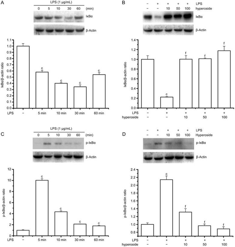
Figure 6.
Effect of hyperoside on LPS-induced phosphorylation and degradation of IκBα. RA FLSs (2×10⁶/mL) were incubated with LPS (1 g/mL) for the indicated time with or without the indicated concentration of hyperoside. In coincubation experiments, hyperoside was added to cells 4 h prior to LPS and then treated with LPS for 30 min (IκBα) or 15 min (phospho-IκBα). The cytosolic extracts of these cells were prepared for Western blotting. (A and B) Representative immunoblot comparing IκBα to β-actin in RA FLSs after the indicated treatment. Group data showing the normalization of IκBα to β-actin as determined in each group from the 3 independent experiments. (C and D) Representative immunoblot comparing phosphorylated IκBα to β-actin in RA FLSs after the indicated treatment. Group data showing the normalization of phosphorylated IκBα to β-actin as determined in each group from the 3 independent experiments. **P<0.01 compared with the untreated control. ##P<0.01 compared with the LPS-treated group.

