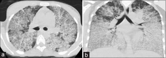
Figure 2.

High-resolution computed tomography chest (a) (lung window) and (b) (coronal section). The anterior part of both the lung fields show typical crazy-paving pattern with central ground-glassing and peripheral interlobular septal thickening. In the dependent part of the lung, there is an increased density secondary to the gravitational accumulation of lipo-proteinaceous fluid
