
Figure 2.
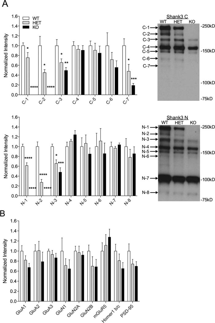
Altered Shank3 isoform expression from whole striatum lysates in Shank3e4–9 mutant mice. (A) Quantification and representative western blots of striatum whole tissue lysates with C-terminal Shank3 antibody (top) and N-terminal Shank3 antibody (bottom) showing decrease (HET) or complete loss (KO) of the C-1, C-2, N-1, and N-2 bands of Shank3 using Shank3 C and N antibodies in Shank3e4–9 mutants compared to WT. Additionally, there was a significant decrease in C-3, C-7, and N-3 bands in both HET and KO mice. (B) Quantification of other synaptic proteins from striatal lysates shows no significant differences. For each analysis, data were normalized to β-actin levels and then to the average of WT (*P < 0.05, **P < 0.01, ***P < 0.001, n = 8 WT, 7 HET, 8 KO).