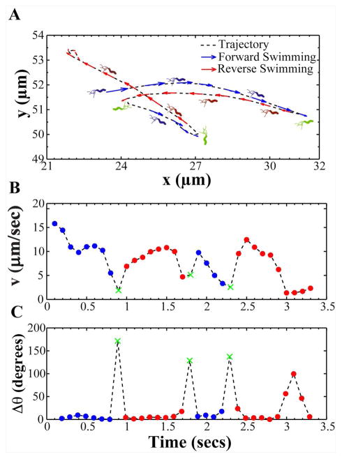
Figure 3. A representative bacterial trajectory depicting the swimming motion of H. pylori.
(A) A representative bacterial trajectory of H. pylori is shown segmented into forward (blue) and reverse swimming directions (red). In our analysis, the direction of swimming observed at the start of the video was considered forward. A reversal in swimming direction (green) was identified when bacteria exhibited a large angle change (Δθ) > 110 degrees. Upon a reversal, bacteria were assumed to continue swimming in the reversed direction (red) until another reversal took place. (B) Instantaneous forward and reversal swimming speeds and (C) change in swimming angle (Δθ) of the bacterium in panel A over the course of time it was tracked (X (green) denotes reversals in swimming direction).

