
Figure 1.
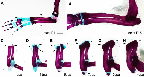
Time course of amputation callus formation in P1 neonatal mice. The whole‐mount samples were stained with alcian blue and alizarin red. (A), (B) Intact forelimbs of P1 (A) and P15 (B) neonates. (C)−(H) Formation of the amputation callus at 1 dpa (C), 3 dpa (D), 5 dpa (E), 7 dpa (F), 10 dpa (G), and 14 dpa (H). The arrowheads indicate the amputation level. The arrows indicate the points where ossification begins in the cartilaginous calluses. Skeletal tissues are indicated; R, radius; U, ulna; Hu, humerus. Scale bar 1 mm (applicable to all photographs).