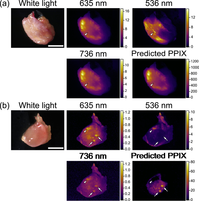
Figure 6. Ex vivo imaging of PPIX fluorescence of representative lymph node metastasis of a colorectal cancer patient.
(a) Metastatic lymph node. (b) Non-metastatic lymph node. Arrowheads indicate unwanted autofluorescence other than PPIX, which was clearly eliminated in the calibrated images of PPIX fluorescence. Arrows show PPIX accumulation in non-metastatic regions, which may indicate follicles. Scale bars in a and b represent 5 mm and 2 mm, respectively.

