
Figure 4.
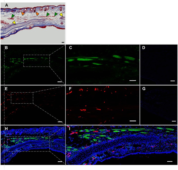
Immunofluorescence analysis of regenerated A. cahirinus ear punch. (A) Masson's trichrome stain (50x); vertical dotted line indicates original plane of wounding; yellow arrowheads indicate cartilage; green arrowheads indicate muscle; orange arrowheads indicate hair follicles. (B) Incubated with anti‐actin (50x). (C) Probed with anti‐actin (200×). (D) Negative control in which the primary antiserum was omitted (200×). (E) Incubated with anti‐TUJ1 (50x). (F) Probed with anti‐TUJ1 (200×). (G) Negative control stained with secondary antibody only (200×). (H) Merge of B + E + Dapi (5x). (I) Merge of B + E + Dapi (200×). All scale bars 100 μm.