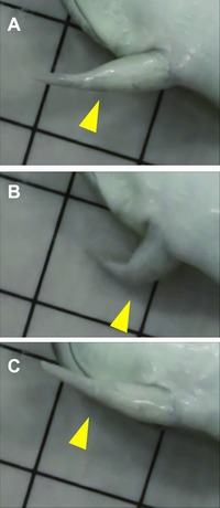
Figure 2.

The bending−stretching motion of the regenerated spike (see also Movie S1). After amputation at the elbow, the motion of the regenerated forelimb was observed. (A) The frog stretched the regenerated forelimb. (B) The frog then bent the regenerated forelimb at the elbow joint. (C) The frog then stretched the limb again. Yellow arrowheads indicate the position of the regenerated elbow between the remaining stylopod and the regenerated spike. The sides of the squares in the background are 1 cm.
