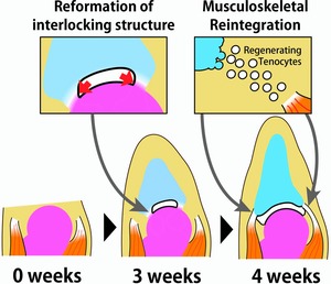
Figure 10.

The process of joint regeneration in frogs. Left panel: Amputated frog forelimb after amputation at the elbow joint. Middle panel: At 3 weeks after amputation, the partial connection between the remaining joint cartilage and the regenerating spike cartilage is divided by a cavity between these apposing cartilages, and the interlocking structure is thereby reformed. Right panel: At 4 weeks after amputation, tenocytes are regenerated between the extremity of the remaining muscles and the regenerating cartilage.
