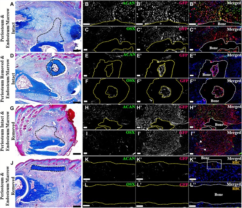
Figure 4.

P2 periosteum and endosteum/marrow contribute to wound repair (dorsal surface is top, distal to the right). (A)−(C′′) Serial sections of an intact GFP‐labeled P2 bone grafted into a NOD‐SCID fractured P2 digit, 11 DPF. Representative sample shown. N = 4. (A) Mallory trichrome staining showing callus formation on the grafted bone (outlined). (B)−(B′′) Double immunostaining for ACAN and GFP illustrating cartilage derived from the bone graft (outlined). (C)−(C′′) Double immunostaining for OSX and GFP showing graft‐derived osteoblasts within the periosteal callus and the endosteal/marrow space (graft outlined). Immunohistochemical stained samples counterstained with DAPI. (D)−(F′′) Serial sections of a periosteum‐removed and endosteum/marrow‐intact GFP‐labeled P2 bone grafted into a NOD‐SCID fractured P2 digit, 11 DPF. Representative sample shown. N = 4. (D) Mallory trichrome staining showing no periosteal callus formation on the grafted bone (outlined). (E)−(E′′) Double immunostaining for ACAN and GFP revealing no graft‐derived chondrocytes present on the periosteal surface or within the marrow cavity (graft outlined). (F)−(F′′) Double immunostaining for OSX and GFP showing double‐labeled osteoblasts present within the graft marrow space (outlined). Immunohistochemical stained samples counterstained with DAPI. (G)−(I′′) Serial sections of periosteum‐intact and endosteum/marrow‐removed GFP‐labeled P2 bone grafted into a NOD‐SCID fractured P2 digit, 11 DPF. Representative sample shown. N = 4. (G) Mallory trichrome staining showing graft periosteal callus formation (outlined). (H)−(H′′) Double immunostaining for ACAN and GFP showing the graft‐derived cartilaginous callus (grafted bone outlined). (I)−(I′′) OSX and GFP double immunostaining showing graft‐derived osteoblasts (arrowheads). Immunohistochemical stained samples counterstained with DAPI. (J)−(L′′) Serial sections of a periosteum and endosteum/marrow‐removed GFP‐labeled P2 bone grafted into a NOD‐SCID fractured P2 digit, 11 DPF. Representative sample shown. N = 4. (J) Mallory trichrome staining showing no callus formation on the grafted bone (outlined). (K)−(K′′) Double immunostaining for ACAN and GFP indicating no graft‐derived chondrocytes present (grafted bone outline). (L)−(L′′) Double labeled osteoblasts were not detected by OSX and GFP double immunostaining (grafted bone outlined). (K)−(L′′) Signal indicates red blood cells (RBC). Immunohistochemical stained samples counterstained with DAPI. Scale bars: (A), (D), (G), (J) 200 μm; all immunostaining 50 μm.
