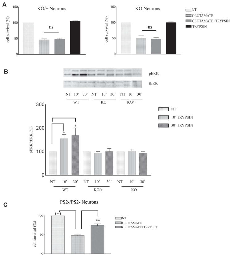
Figure 3. PS1 specifically promotes trypsin-induced neuroprotection and ERK1/2 phosphorylation.
(A) Seven DIV mouse PS1 KO/+ (left) and PS1 KO (right) neuronal cultures were treated with 5.25 nM trypsin for 60 minutes before 3h exposure to glutamate (50μM) as described in Fig. 1. Cell survival assay showed that absence of one PS1 allele is sufficient to inhibit trypsin-mediated neuroprotection. Results (mean ± standard error [SE]) were calculated from 4 independent experiments. (B) (Upper) WT, PS1 KO/+ and PS1 KO neuronal cultures prepared as in Fig. 1 from the same littermate were treated at 7 DIV with 5.25 nM trypsin for the indicated time periods. Untreated cultures (NT) were used as controls. Following incubation, cells were collected and assayed on WB for the indicated proteins. A representative blot out of 5–7 independent experiments is shown. (Lower) Densitometric analysis of p-ERK 1/2 in the presence of trypsin at different time points expressed as percentage of phospho-ERK 1/2 (pERK) to total ERK 1/2 (t-ERK) ratio that was set as 100% for control (NT). Bars represent phospho-protein to total protein ratios relative to control. *p=0.05, ** p<0.005 (Tukey’s post-hoc). (C) PS2 −/− 7DIV neuronal cultures were treated with 5.25nM trypsin 60 minutes prior to the addition of 50 μM glutamate for 3h. Cell survival assay shows that trypsin rescued cortical neurons from glutamate excitotoxicity in the absence of PS2, n=3 **p<0.005, *** p<0.001 (Tukey’s post hoc).

