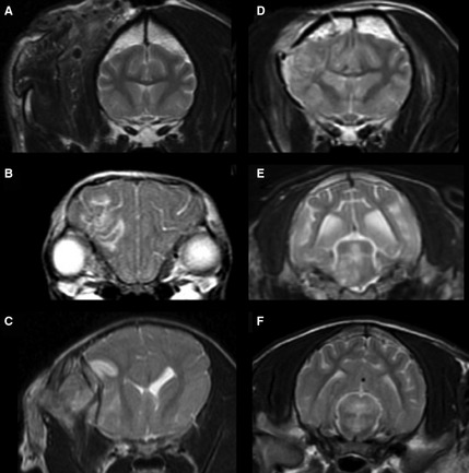
Figure 1.

Brain magnetic resonance imaging (MRI) of dogs with traumatic brain injury. Transverse T2‐weighted images Fast Spin Echo (A–F) showing the MRI grading system. (A) Grade I—normal parenchyma. (B) Grade II—lesions only affecting the cerebral hemisphere, cerebellar parenchyma without midline shift or both. (C) Grade III—lesions only affecting the cerebral hemisphere, cerebellar parenchyma, or both and causing midline shift. (D) Grade IV—lesions affecting corpus callosum, thalamus, or basal nuclei with or without any of the foregoing lesions of lesser grades. (E) Grade V—unilateral lesions in the brainstem with or without any of the foregoing lesions of lesser grades. (F) Grade VI—bilateral lesions affecting the brainstem with or without any of the foregoing lesions of lesser grades.
