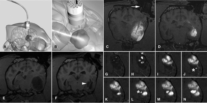
Figure 10.

Convection‐enhanced delivery of liposomal CPT‐11 (a topoisomerase inhibitor) intratumorally by using real‐time MR imaging to optimize delivery. (A, B) Schematic representation of fused silica cannulae being guided into the tumor based on stereotactically placed guide pedestals. (C, D) Transverse T1‐weighted images at different levels showing infusate (white) of liposomal CPT‐11 and gadoteridol contrast agent within the tumor. Different cannulae can be seen highlighted against the infusate after passing down the guide pedestal (arrow). (E, F) Tumor volume (hypointense) pre‐ and posttreatment is decreased by 90% (arrowhead) after CPT‐11 infusion. (G–N) Time‐lapse imaging over approximately 2 hours infusion. Three initial cannulae result in partial tumor coverage. Real‐time imaging allows monitoring of infusion, and placement of additional cannulae (*) resulting in optimal volume of coverage.
