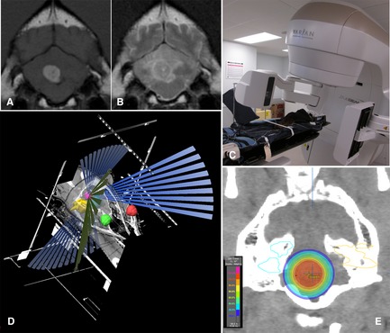
Figure 11.

(A, B) T1‐weighted postcontrast and T2‐weighted transverse images of a 4th ventricle choroid plexus tumor. (C) Dog positioned in stereotactic thermoplastic head restraint. The VARIAN trueBEAM linear accelerator11 is equipped with a 2.5‐mm leaf multileaf collimator, a couch with 0.1‐mm incremental movement, and on‐board Kv, MV and cone beam CT to allow precise stereotactic delivery of radiation. (D) BrainLab planning system10 showing the planned treatment trajectories to the tumor (magenta) sparing defined vital structures (eyes‐red/green, inner ears‐yellow/blue). (E) Transverse CT image with isodose planning superimposed (images courtesy of M. Kent, UC Davis).
