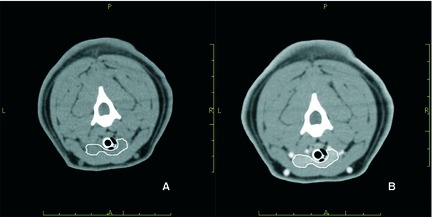
Figure 2.

Transverse precontrast (A) and postcontrast (B) computed tomographic images of the neck. The thyroid tissue (outlined in white) is isoattenuating to the surrounding musculature in the precontrast image (A) and is hyperattenuating in the postcontrast image (B).
