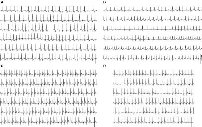
Figure 2.

Electrocardiogram (ECG) from 3 small breed dogs with advanced myxomatous mitral valve disease during a syncopal episode. The ECG strips show 75 seconds of a single precordial lead from the 24‐hour Holter recording displayed chronologically and centered around a syncopal episode. (A) Jack Russell Terrier having a syncopal episode showing sinus rhythm at HR = 131 bpm. (B) Dachshund having sinus rhythm with a shift in HR from 126 to 181 bpm. (C) and (D) ECG showing 2 syncopal episodes from a Miniature Poodle with sinus rhythm at HR 171 and 136 bpm, respectively. bpm, beats per minute; HR, heart rate.
