
Figure 1.
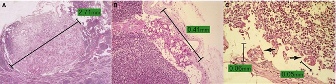
(A) Neoplastic proliferations characterized by numerous structures resembling acini contain significant fibrosis replacing part of the lymphoid parenchyma (macrometastases). 400x. Hematoxylin‐eosin (H&E). (B) Neoplastic proliferation characterized by structures resembling acini localized in the subcapsular sinus (micrometastases). 200x. H&E. (C) Multiple deposits of isolated epithelial neoplastic cells (arrows) in the subcapsular sinus. The size of the largest metastatic deposit, measuring 0.06 mm, was used to classify lymph node metastasis as isolated tumor cells (ITC). 400x. H&E.