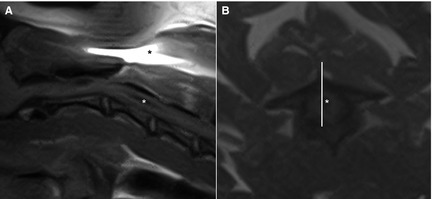
Figure 2.

Midsagittal (A) and transverse (B) T1‐weighted spin echo image of the cranial cervical spine of the same dog. A hypointense cavity (white asterisk) is visible within the spinal cord. A susceptibility artifact (*) is visible because of the presence of a microchip. The corresponding slice (white line) of the sagittal image is off midline compared to the transverse image.
