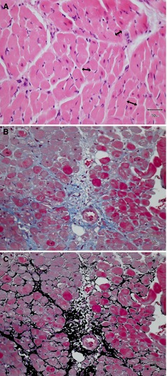
Figure 3.

Histologic morphometry of the myocardium by digitization at original magnification (×40). (A) Diameter of myocardium was randomly measured at the level of nucleus (double‐headed arrows), (B) Representative Masson's trichrome stained myocardial section for the quantification of the interstitial fibrous tissue, (C) The same photograph of (B) to illustrate the area of the interstitial connective tissue transformed by computer‐based pixel data.
