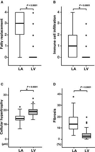
Figure 5.

Histopathologic disparity between the LA and the LV. The fatty replacement (A), the immune cell infiltration (B), and the extent of the fibrosis (D) were significantly higher in the LA than in the LV. In particular, the values of the fatty replacement and the inflammatory cell infiltration were nearly zero in the LV. The size of myocytes was significantly larger in the LV than in the LA, indicating cellular hypertrophy (C).
