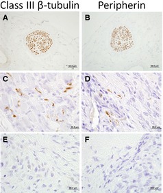
Figure 5.

Microscopic appearance of a peripheral sensory nerve traversing through control tissue as indicated by positive immunohistochemical staining for (A) class III β tubulin and (B) peripherin. Haphazard and disorganized proliferation of neuronal endings identified within OS‐associated periosteum as determine by positive immunohistochemical staining for (C) class III β tubulin and (D) peripherin. Representative OS‐associated periosteum which is devoid of excessive neuronal end proliferations based upon the absence of (E) class III β tubulin and (F) peripherin immunohistochemical staining.
