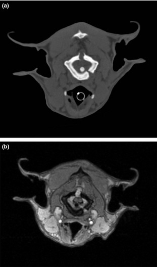
Figure 1.

Transverse CT images of one of the Czechoslovakian wolfdog dwarfs at the level of C1 (a). Note the open suture lines of C1. Transverse postcontrast flash 3D‐WI of the same dog at the level of C1 (b). Note the enhancement of the tissue in the dorsal and left ventral suture. Note the dorsal displacement of the spinal cord, which is mildly compressed between the dens and the suture tissue.
