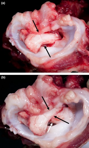
Figure 2.

Transverse cranial macroscopic view of the cervical vertebral column including C1, at necropsy of a Czechoslovakian wolfdog with dwarfism. (a) Extension of C1‐C2. There is no compression of the spinal cord (black arrows). (b) Flexion of C1‐C2. Note the compression of the cervical spinal cord (black arrows) caused by dorsal displacement of the dens axis (white arrow).
