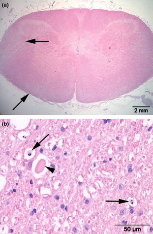
Figure 4.

Transverse section of the cervical spinal cord at C1 (a) revealing vacuolar changes in ventral and lateral funiculi (arrows). Marked axonal swelling (arrowhead) and influx of macrophages (arrows) consistent with Wallerian degeneration (b). H&E.
