
Figure 1.
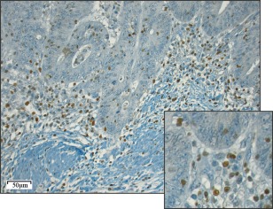
T‐bet immunoreactivity in CRC tumour specimens. Representative light microscopic image of immunohistochemical staining for T‐bet in a CRC specimen (20 objective magnification). The smaller square represents a close‐up using 40 objective magnification.