
Figure 2.
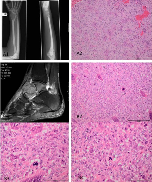
Photomicrographs and X‐rays of a conventional (S00033682) (A), and malignant giant cell tumour (GCT) (S00030176) (B). A central lytic tumour of the distal radius (A1). The histology is typical for GCT in which the stromal cells show no significant cytological atypia (A2). An aggressive lytic lesion of the talus, which has broken through the cortex (B1). Microscopy shows features of a malignant osteoclast‐rich tumour in which the stromal cell are enlarged with nuclear pleomorphism and numerous mitoses, including atypical forms (B3 and B4). Geographic‐type tumour necrosis was also present (not shown).