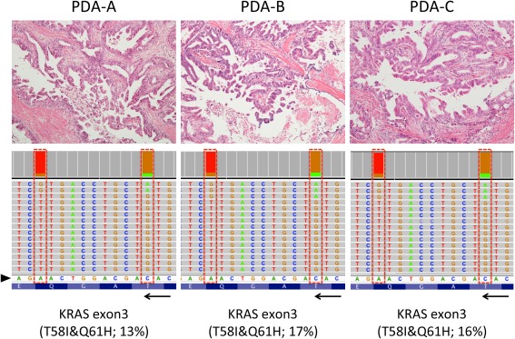
Figure 1.

Representative histology and KRAS mutation pattern in three metachronous PDAs. Histologies of the three PDAs are shown (top panels; original magnifications, ×10 objective). All three tumours were moderately differentiated adenocarcinoma with papillary proliferation of cancer cells. Each sample shows the representation of the reads aligned to the reference genome in the KRAS genes (displayed as reverse‐complement of Exon 3; bottom panels). Arrowhead and arrow indicate of forward sequences of KRAS gene and its direction (3′ < 5′). Those histologically similar lesions possessed unique haplotype in KRAS (p.T58I&p.Q61H) analysed separately using NGS.
