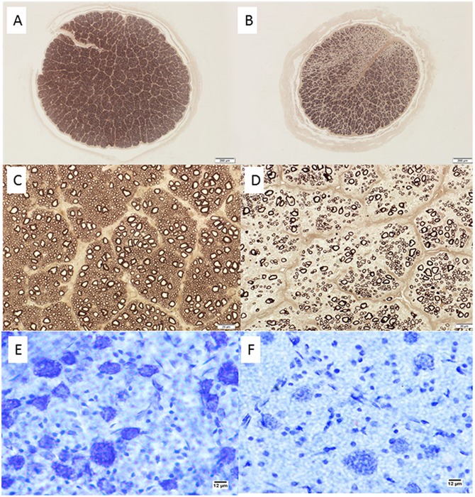
Fig 6. Retinal Ganglion Cell Loss in Cats With Primary Congenital Glaucoma.
Photomicrographs showing representative optic nerve sections [A-D], stained with p-phenylenediamine to highlight axonal myelin sheaths, from 2 young adult cats. [A,C] normal cat (estimated 83,398 optic nerve axons); [B,D] typical PCG-affected cat with moderate degree of axon loss (estimated 30,365 optic nerve axons). Mid-peripheral retina in cresyl violet stained retinal whole mounts from these same two cats show relative loss of Nissl-stained retinal ganglion cell (RGC) somas of >12μm diameter in the ganglion cell layer of the retina in PCG (total RGC soma count in whole retina of 43,891)[E] relative to a normal cat [F] (estimated RGC soma count in whole retina of 123,833). Bar markers = 200μm (top row); 20μm (middle row), and 12μm (bottom row).

