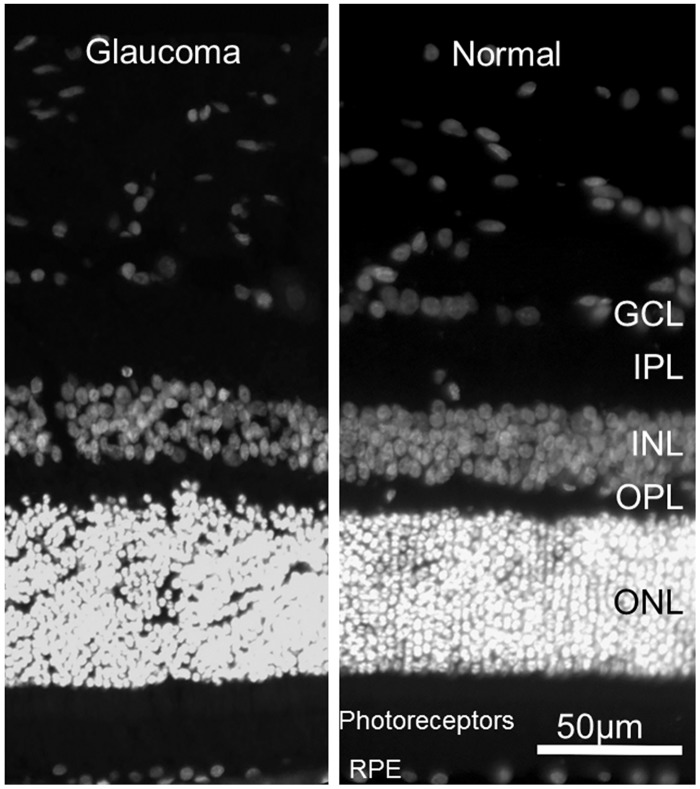
Fig 8. Preservation of Normal Outer Retinal Structure in Feline Primary Congenital Glaucoma.
Representative fluorescence photomicrographs of DAPI stained 18 month old glaucomatous [A] and normal 6 month old [B] feline retinas. No significant difference in morphology of outer retinal layers is identified. (GCL = ganglion cell layer; IPL = inner plexiform layer; INL = inner nuclear layer; OPL = outer plexiform layer; ONL = outer nuclear layer; RPE = retinal pigment epithelium).

