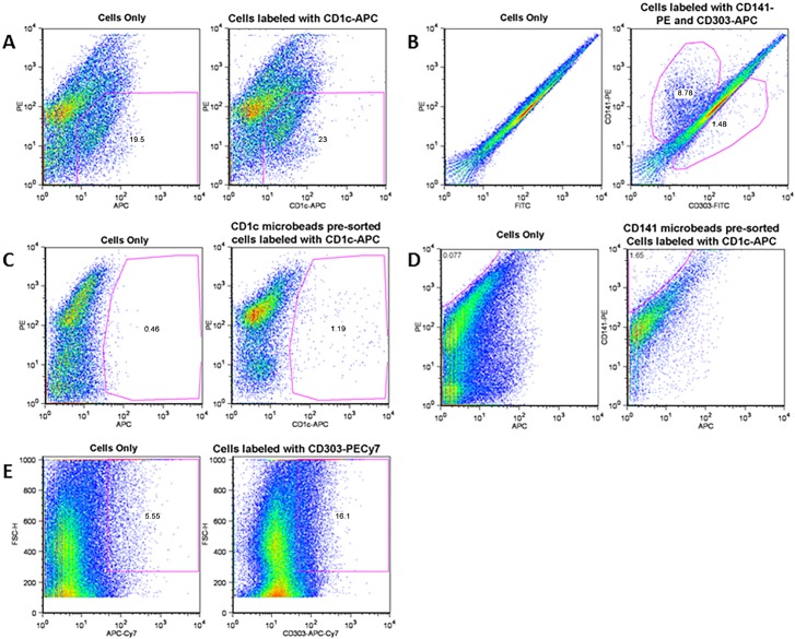
Fig 5. Dynamics of myeloid and plasmacytoid dendritic cells in S and AS carotid plaques.
Cell characterization was performed using anti-human antibodies for mDC1 (CD1c+), mCD2 (CD141+) and pDC (CD303+). (A) Cells only are without antibody (background) and cells labeled with CD1C antibody for mDC1. After subtracting background, mDC1 cells were 3.5%; (B) CD141+ cells (mDC2) were 8.78% and CD303+ cells (pDCs) were 1.41%; and (C) Cells only are without antibody (background) and cells labeled with CD1C antibody for mDC1. After subtracting background, (D) mDC1 cells were 0.73% and CD141+ mDC2 were 1.58%, and (E) CD303+ cells (pDCs) were 10.7%. The data represent 4 plaques from independent patients in each experimental group.

