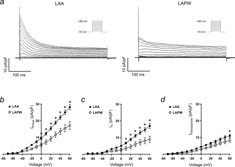
Fig 6. Ito reduction in cardiomyocytes isolated from the left atrial poisterior wall (LAPW).
(a) Example voltage-sensitive, Ca2+ independent, macroscopic K+ currents evoked in cardiomyocytes isolated from the left atrial appendage (LAA, left) and LAPW (right). Voltage protocol is shown inset. (b-d) LAA and LAPW I/V relationships for the peak outward K+ current, Ito and steady state K+ current. Data presented as mean ± SEM. * denotes P<0.05 LAA (N = 16 cells) v LAPW (N = 12 cells), two way repeated measures Analysis of Variance (ANOVA) with Bonferroni post hoc analysis.

