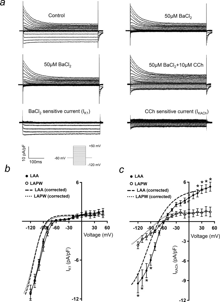
Fig 7. IKACh is depleted in left atrial posterior wall (LAPW) cardiomyocytes.
(a) Current traces demonstrating isolation of BaCl2 sensitive (IK1) and CCh induced (IKACh) currents in a single left atrial cardiomyocyte. Voltage protocol is shown inset. (b & c) Comparison of LAA and LAPW I/V relationships for IK1 and IKACh. The dashed lines indicate mean best fit IK1 and IKACh I/V curves with liquid junction potential correction, for both LAA and LAPW. Data presented as mean ± SEM. * denotes P<0.05 LAA (N = 25 cells) v LAPW (N = 17 cells), two way repeated measures Analysis of Variance (ANOVA) with Bonferroni post hoc analysis.

